
Lower-extremity diabetic ulcers are responsible for 80% of annual worldwide nontraumatic amputations. Epidermal growth factor (EGF) reduction is one of the molecular pillars of diabetic ulcer chronicity, thus EGF administration may be considered a type of replacement therapy. Topical EGF administration to improve and speed wound healing began in 1989 on burn patients as part of an acute-healing therapy. Further clinical studies based on topically administering EGF to different chronic wounds resulted in disappointing outcomes. An analysis of the literature on unsuccessful clinical trials identified a lack of knowledge concerning: (I) molecular and cellular foundations of wound chronicity and (II) the pharmacodynamic requisites governing EGF interaction with its receptor to promote cell response. Yet, EGF intra- and perilesional infiltration were shown to circumvent the pharmacodynamic limitations of topical application. Since the first studies, the following decades of basic and clinical research on EGF therapy for problem wounds have shed light on potential uses of growth factors in regenerative medicine. EGF’s molecular and biochemical effects at both local and systemic levels are diverse: (1) downregulation of genes encoding inflammation mediators and increased expression of genes involved in cell proliferation, angiogenesis and matrix secretion; (2) EGF intervention positively impacts both mesenchymal and epithelial cells, reducing inflammation and stimulating the recruitment of precursor circulating cells that promote the formation of new blood vessels; (3) at the subcellular level, upregulation of the EGF receptor with subsequent intracellular trafficking, including mitochondrial allocation along with restored morphology of multiple organelles; and (4) local EGF infiltration resulting in a systemic, organismal repercussion, thus contributing to attenuation of circulating inflammatory and catabolic reactants, restored reduction-oxidation balance, and decreased toxic glycation products and soluble apoptogenic effectors. It is likely that EGF treatment may rearrange critical epigenetic drivers of diabetic metabolic memory.
KEYWORDS Epidermal Growth Factor, diabetes, diabetes complications, wound healing, diabetic foot, amputation, ulcer, Cuba
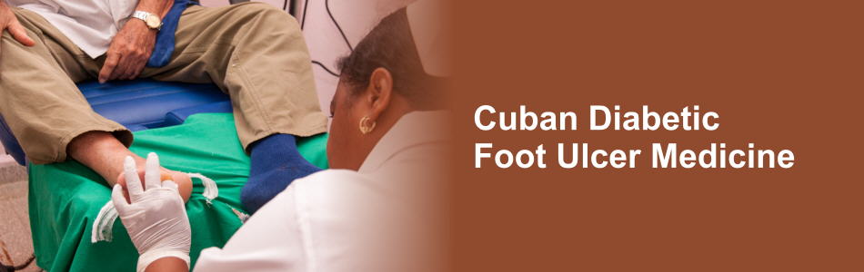
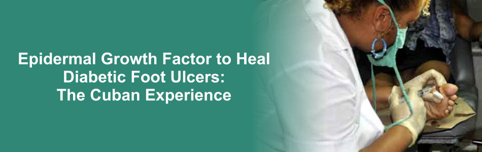
INTRODUCTION Diabetic foot ulcers are a chronic complication in patients with diabetes mellitus. They appear as a result of the combination of diabetic polyneuropathy and angiopathy, and in many cases require amputation of the affected extremity. Clinical trials have demonstrated that repeated local infiltration with Heberprot-P can improve healing of chronic diabetic foot ulcers. Although there is evidence of its effects as a granulation stimulator and on cell migration and proliferation, genetic control mechanisms explaining its anti-inflammatory and oxidative stress reduction properties are not yet thoroughly understood.
OBJECTIVE Analyze changes in expression of genes involved in healing after treatment of diabetic foot ulcers with Heberprot-P.
METHODS Biopsies were collected from diabetic foot ulcers of 10 responding patients before and after 2 weeks’ treatment with Heberprot-P (75-μg applied intralesionally 3 times per week). Total RNA was obtained and quantitative PCR used to determine expression of 26 genes related to inflammation, oxidative stress, cell proliferation, ngiogenesis and extracellular matrix formation. Genetic expression was quantified before and after treatment using REST 2009 v2.0.13.
RESULTS After treatment, there was a statistically significant increase in expression of genes related to cell proliferation, angiogenesis and formation of extracellular matrix (PDGFB, CDK4, P21, TP53, ANGPT1, COL1A1, MMP2 and TIMP2). A significant decrease was observed in gene expression related to inflammatory processes and oxidative stress (NFKB1, TNFA and IL-1A).
CONCLUSIONS Our findings suggest that Heberprot-P’s healing action on diabetic foot ulcers is mediated through changes in genetic expression that reduce hypoxia, inflammation and oxidative stress, and at the same time increase cell proliferation, collagen synthesis and extracellular matrix remodeling. The kinetics of expression of two genes related to extracellular matrix formation needs further exploration.
KEYWORDS Epidermal growth factor, EGF, diabetic foot ulcer, wound healing, quantitative real-time PCR, gene expression, Cuba
